
Tissue engineering has been revolutionized since 2004 by the emergence of three-dimensional (3D) bioprinting.[1] In this approach, additive manufacturing techniques (microextrusion, laser-assisted printing, and inkjet printing) are used to create autologous biological tissues. Mesenchymal stem cells or differentiated cells are placed in a fluid hydrogel to form a "bio-ink."[2]
The computer-assisted design of the numerical 3D model is based on an ".stl" file, which can be produced from the computed tomography scan of a tissue defect.[3]
To date, the tissues (eg, liver tissue, heart tissue, skin, cartilage) produced with in vitro 3D bioprinting have mostly been centimeter scale, and promising results have been obtained. In vitro bioprinting of composite tissues with different cell types has also been performed with a few encouraging results in animal studies.[4] The growing interest in 3D bioprinting is highlighted by the tripling between 2012 and 2015 of research articles published on this topic. Four journals dedicated specifically to bioprinting have recently been launched.
The 3d.FAB unit at the University of Lyon (Université Claude Bernard-Lyon 1), created in early 2015, is a unique platform in Europe in which biochemists, biologists, engineers, and physicians are studying applications for 3D printing in medicine and the life sciences, notably bioprinting (Web site: http://fabric-advanced-biology.univ-lyon1.fr/).
Our team, in partnership with LabSkin Creations and the Laboratory of Cutaneous Substitutes (Edouard Herriot Hospital, Lyon) has developed a specific hydrogel for skin microextrusion bioprinting. Its composition has been patented. We routinely prepare skin in reproducible 1-cm2 pieces, 0.5-cm thick (Fig. 1), and pieces as large as 200 cm2 on demand. Histological examination shows that the skin samples prepared are of high quality (Fig. 2). Cell viability is 100%. Using this approach, full-thickness skin is obtained within 21 days, compared with 45 days for traditional tissue engineering techniques.
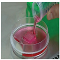
Figure 1.
Photograph of a skin piece at the end of microextrusion process.

Figure 2.
Comparison of optical microscopy images of equivalent slices of normal human skin and printed skin after 26 days of culture. The tissues were stained with Masson's trichrome. DEJ, dermoepidermal junction.
We will focus next on the bioprinting of bone. Three different types of bioprinted bone grafts for mandibular reconstruction will be compared, first in vitro and then after implantation in animals.
The unique characteristic of 3D bioprinting is the cellular ink that is used, from which precisely shaped grafts can be produced. This technique differs from traditional tissue engineering in which a scaffold is first created and then filled with cells.
On a microscopic scale, 3D bioprinting allows a cell-specific environment to be created with a conducive 3D structure. Cells are dispersed in the hydrogel, which contains all the extracellular factors required for their survival.
On a macroscopic scale, the main advantage of 3D bioprinting is that it will allow the creation of personalized autologous grafts that perfectly fit the tissue defect and thereby increase the success of the reconstruction.
Autologous grafts produced by computer-assisted manufacturing will be faster to implant and minimize donor site morbidity. The practical limitation in terms of graft size is the ability to create a vascular network. Once this limit is overcome, the production of "ready-to-implant" organs and autologous free flaps with a vascular pedicle will become possible.
Plast Reconstr Surg Glob Open. 2017;5(3):e1246 © 2017 Lippincott Williams & Wilkins


