Practice Essentials
Diabetic foot ulcers, as shown in the images below, occur as a result of various factors, such as mechanical changes in conformation of the bony architecture of the foot, peripheral neuropathy, and atherosclerotic peripheral arterial disease, all of which occur with higher frequency and intensity in the diabetic population. [1, 2]
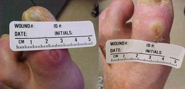 Diabetic ulcer of the medial aspect of left first toe before and after appropriate wound care.
Nonenzymatic glycation predisposes ligaments to stiffness. Neuropathy causes loss of protective sensation and loss of coordination of muscle groups in the foot and leg, both of which increase mechanical stresses during ambulation.
Diabetic foot lesions are responsible for more hospitalizations than any other complication of diabetes. [3] Diabetes is the leading cause of nontraumatic lower extremity amputations in the United States, with approximately 5% of diabetics developing foot ulcers each year and 1% requiring amputation.
Physical examination of the extremity that has a diabetic ulcer can be divided into examination of the ulcer, examination of the feet, assessment of the possibility of vascular insufficiency, [4] and assessment for the possibility of peripheral neuropathy.
Diabetic foot ulcers can be staged using the Wound, Ischemia, and foot Infection (WIfI) threatened limb classification system. This system allows communication between providers and provides risk stratification for major amputation. [5] Blood work should be obtained, such as a complete blood count (CBC), a comprehensive metabolic panel, and hemoglobin A1c (HbA1c), as well as inflammatory markers when infection is suspected. Weight-bearing radiographs of the affected limb should be obtained.
The management of diabetic foot ulcers requires offloading the wound, [6, 7] daily saline or similar dressings to provide a moist wound environment, [8] débridement when necessary, antibiotic administration with or without surgical intervention if osteomyelitis or soft tissue infection is present, [9, 10] optimal control of blood glucose, and evaluation and correction of peripheral arterial insufficiency. [11]
All patients harboring diabetic foot ulcers should be evaluated by a qualified vascular surgeon and podiatric surgeon who will consider débridement, reconstructive surgery on bony architecture, vascular reconstruction, and options for soft tissue coverage.
It is prudent to address the underlying etiologies in diabetic foot ulcers for wound care modalities to be successful. Without addressing the osseous deformities and muscular imbalances, infections, and vascular insufficiency, there will be of minimal benefit in employing advanced wound care dressings.
For more information, see Diabetes Mellitus, Type 1 and Diabetes Mellitus, Type 2.
Pathophysiology
Atherosclerosis and peripheral neuropathy occur with increased frequency in persons with diabetes mellitus (DM).
Trophic changes
Non-enzymatic glycosylation of skin and connective tissue, along with decreased collagen production in people with diabetes, result in alterations in the biomechanics in the diabetic foot. This is frequently seen in the Achilles tendon, where increased stiffness results in a contracture that limits ankle dorsiflexion, a condition known as equinus. Equinus has been associated with diabetic foot ulcers, as it increases plantar pressures in the forefoot and midfoot.
Diabetes-related atherosclerosis
Overall, people with diabetes mellitus (DM) have a higher incidence of atherosclerosis, thickening of capillary basement membranes, arteriolar hyalinosis, and endothelial proliferation. Calcification and thickening of the arterial media (Mönckeberg sclerosis) are also noted with higher frequency in the diabetic population, although whether these factors have any impact on the circulatory status is unclear.
Diabetic persons, like people who are not diabetic, may develop atherosclerotic disease of large-sized and medium-sized arteries, such as aortoiliac and femoropopliteal atherosclerosis. However, significant atherosclerotic disease of the infrapopliteal segments is particularly common in the diabetic population. Underlying digital artery disease, when compounded by an infected ulcer in close proximity, may result in complete loss of digital collaterals and precipitate gangrene.
The reason for the prevalence of this form of arterial disease in diabetic persons is thought to result from a number of metabolic abnormalities, including high low-density lipoprotein (LDL) and very-low-density lipoprotein (VLDL) levels, elevated plasma von Willebrand factor, inhibition of prostacyclin synthesis, elevated plasma fibrinogen levels, and increased platelet adhesiveness.
Diabetic peripheral neuropathy
The pathophysiology of diabetic peripheral neuropathy is multifactorial and is thought to result from vascular disease occluding the vasa nervorum; endothelial dysfunction; deficiency of myoinositol-altering myelin synthesis and diminishing sodium-potassium adenine triphosphatase (ATPase) activity; chronic hyperosmolarity, causing edema of nerve trunks; and effects of increased sorbitol and fructose. [12]
Motor dysfunction of peripheral nerves in diabetic neuropathy leads to muscular imbalances in the diabetic foot. Muscle wasting of the intrinsic pedal muscles leads to overpowering of the spared extrinsic muscles, which results in significant forefoot deformities such as claw toes or hammer toes. [13, 14] Autonomic dysfunction of the peripheral nervous system may lead to sudomotor dysfunction. This will result in dry, cracked skin, which is more prone to injury and breakdown. [15]
The result of loss of sensation in the foot is repetitive stress; unnoticed injuries and fractures; structural foot deformity, such as hammertoes, bunions, metatarsal deformities, or Charcot foot (see the image below); further stress; and eventual tissue breakdown. Unnoticed excessive heat or cold, pressure from a poorly fitting shoe, or damage from a blunt or sharp object inadvertently left in the shoe may cause blistering and ulceration. These factors, combined with poor arterial inflow, confer a high risk of limb loss on the patient with diabetes.
See Diabetic Neuropathy for more information.
Etiology
The etiologies of diabetic ulceration include neuropathy, [16] arterial disease, [17] pressure, [6] and foot deformity. [18] Diabetic peripheral neuropathy, present in 60% of diabetic persons and 80% of diabetic persons with foot ulcers, confers the greatest risk of foot ulceration; microvascular disease and suboptimal glycemic control contribute.
A study by Naemi et al indicated that tissue mechanics may be associated with foot ulceration in patients with diabetic neuropathy, with an evaluation of 39 patients finding that the heel pad in nonulcerated feet tended to be stiffer than in ulcerated feet. [19] . These results were further elucidated in another study by Naemi et al, which reported that the risk of diabetic foot ulcer is higher in diabetic neuropathy patients who have greater plantar soft tissue thickness and lower stiffness in the area of the first metatarsal head. The investigators found that adding the mechanical properties of plantar soft tissue (stiffness and thickness) to commonly evaluated clinical parameters improved specificity, sensitivity, prediction accuracy, and prognosis strength by 3%, 14%, 5%, and 1%, respectively. [20]
The anatomy of the foot must be considered in risk calculation. A person with flatfoot is more likely to have disproportionate stress across the foot and may have an increased risk for tissue inflammation in high-stress regions.
Charcot foot
Sensory neuropathy involving the feet may lead to unrecognized episodes of trauma due to ill-fitting shoes. Motor neuropathy, causing intrinsic muscle weakness and splaying of the foot on weight bearing, compounds this trauma. The result is a convex foot with a rocker-bottom appearance. Multiple fractures are unnoticed until bone and joint deformities become marked. This is termed a Charcot foot (neuropathic osteoarthropathy) and most commonly is observed in diabetes mellitus, affecting about 2% of diabetic persons.
If a Charcot foot is neglected, ulceration may occur at pressure points, particularly the medial aspect of the navicular bone and the inferior aspect of the cuboid bone. Sinus tracts progress from the ulcerations into the deeper planes of the foot and into the bone. Charcot change can also affect the ankle, causing displacement of the ankle mortise and ulceration, which can lead to the need for amputation.
Epidemiology
According to the National Institute of Diabetes and Digestive and Kidney Diseases, an estimated 16 million Americans are known to have diabetes, and millions more are considered to be at risk for developing the disease. Diabetic foot lesions are responsible for more hospitalizations than any other complication of diabetes. [3] Among patients with diabetes, there is a 25% likelihood that they will develop a foot ulcer. [21] Nearly half of them will become infected, and about 20% of those with moderate to severe infection will require some degree of amputation. [22, 23] Indeed, diabetes is the leading cause of nontraumatic lower extremity amputations, as it accounts for 85% of these procedures. [21]
Mortality
People with diabetes who have ulcerations are at 2.5 times higher risk of death at 5 years, versus those without ulcerations. [24] While patients who have had a diabetes-related amputation have a 70% risk of mortality at 5 years, those on dialysis have an even greater risk, at 74%. [25]
Age distribution for diabetic ulcers
Diabetes occurs in 3-6% of Americans. Of these, 10% have type 1 diabetes and are usually diagnosed when they are younger than 40 years. Among Medicare-aged adults, the prevalence of diabetes is about 10% (of these, 90% have type 2 diabetes). Diabetic neuropathy tends to occur about 10 years after the onset of diabetes, and, therefore, diabetic foot deformity and ulceration occur sometime thereafter.
Prevalence of diabetic ulcers by race
The issue of diabetic foot disease is of particular concern in the Latino communities of the Eastern United States, in African Americans, [26] and in Native Americans, who tend to have the highest prevalence of diabetes in the world.
See Diabetic Foot Infections for more information.
Prognosis
Mortality in people with diabetes and foot ulcers is often the result of associated large vessel arteriosclerotic disease involving the coronary or renal arteries.
In assessing the health-related quality of life (HRQOL) in adults with diabetic foot ulcers, a literature review by Khunkaew et al found that such patients scored poorly on four of eight scales on the 36-Item Short Form Health Survey (SF-36), specifically, physical functioning, role physical, general health, and vitality. Risk factors for a lower HRQOL included the existence of pain, a C-reactive protein level above 10 mg/L, an ulcer size of over 5 cm2, an ankle-brachial index value of less than 0.9, a high glycosylated hemoglobin level, and a body mass index of over 25 kg/m2. [27]
Limb loss is a significant risk in patients with diabetic foot ulcers, particularly if treatment has been delayed. [28] Diabetes is the predominant etiology for nontraumatic lower extremity amputations in the United States. Half of all nontraumatic amputations are a result of diabetic foot complications, and the 5-year risk of needing a contralateral amputation is 50%. [29]
In diabetic people with neuropathy, [30] even if successful management results in healing of the foot ulcer, the recurrence rate is 66% and the amputation rate rises to 12%.
A study by Chammas et al indicated that ischemic heart disease is the primary cause of premature death in patients with diabetic foot ulcer, finding it to be the major source of mortality on postmortem examination in 62.5% of 243 diabetic foot ulcer patients. The study also found that in patients with diabetic foot ulcer, the mean age of death from ischemic heart disease, as derived from postmortem examination, was 5 years below that of controls. Patients with neuropathic foot ulcers were determined to have the highest risk of premature death from ischemic heart disease. [31]
A study by Chen et al indicated that following hospital treatment for diabetic foot ulcer, invasive systemic infection associated with the ulcer (DFU-ISI) is an important late complication that increases mortality risk. In the study’s patients, methicillin-resistant Staphylococcus aureus (MRSA) gave rise to 57% of the ISIs. Using Cox regression modeling, the investigators found that complicated ulcer healing and the presence of MRSA in the initial ulcer culture predicted the development of DFU-ISIs (hazard ratios of 3.812 and 2.030, respectively), with the hazard ratio for mortality risk in association with DFU-ISIs being 1.987. [32]
Patient Education
The risk of foot ulceration and limb amputation in people with diabetes is lessened by patient education stressing the importance of routine preventive foot care, daily self inspection of feet, appropriate shoes, avoidance of barefoot walking, avoidance of cigarette smoking, control of hyperlipidemia, and adequate glycemic control.
-
Diabetic ulcer of the medial aspect of left first toe before and after appropriate wound care.
-
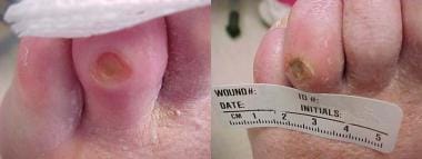
Diabetic ulcer of left fourth toe associated with mild cellulitis.
-
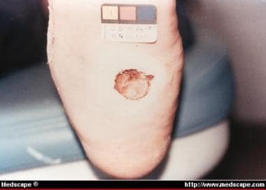
Charcot deformity with mal perforans ulcer of plantar midfoot.
Tables
Category |
Examples |
Description |
Applications |
Alginate |
AlgiSite Comfeel Curasorb Kaltogel Kaltostat Sorbsan Tegagel |
This seaweed extract contains guluronic and mannuronic acids that provide tensile strength and calcium and sodium alginates, which confer an absorptive capacity. Some of these can leave fibers in the wound if they are not thoroughly irrigated. These are secured with secondary coverage. |
These are highly absorbent and useful for wounds having copious exudate. Alginate rope is particularly useful to pack exudative wound cavities or sinus tracts. |
Hydrofiber |
Aquacel Aquacel-Ag Versiva |
An absorptive textile fiber pad, also available as a ribbon for packing of deep wounds. This material is covered with a secondary dressing. The hydrofiber combines with wound exudate to produce a hydrophilic gel. Aquacel-Ag contains 1.2% ionic silver that has strong antimicrobial properties against many organisms, including methicillin-resistant Staphylococcus aureus and vancomycin-resistant Enterococcus. |
These are absorbent dressings used for exudative wounds. |
Debriding agents |
Hypergel (hypertonic saline gel) Santyl (collagenase) Accuzyme (papain urea) |
Various products provide some degree of chemical or enzymatic débridement. |
These are useful for necrotic wounds as an adjunct to surgical débridement. |
Foam |
LYOfoam Spyrosorb Allevyn |
Polyurethane foam has some absorptive capacity. |
These are useful for cleaning granulating wounds having minimal exudate. |
Hydrocolloid |
Aquacel CombiDERM Comfeel Duoderm CGF Extra Thin Granuflex Tegasorb |
These are made of microgranular suspension of natural or synthetic polymers, such as gelatin or pectin, in an adhesive matrix. The granules change from a semihydrated state to a gel as the wound exudate is absorbed. |
They are useful for dry necrotic wounds, wounds having minimal exudate, and clean granulating wounds. |
Hydrogel |
Aquasorb Duoderm IntraSite Gel Granugel Normlgel Nu-Gel Purilon Gel (KY jelly) |
These are water-based or glycerin-based semipermeable hydrophilic polymers; cooling properties may decrease wound pain. These gels can lose or absorb water depending upon the state of hydration of the wound. They are secured with secondary covering. |
These are useful for dry, sloughy, necrotic wounds (eschar). |
Low-adherence dressing |
Mepore Skintact Release |
These are various materials designed to remove easily without damaging underlying skin. |
These are useful for acute minor wounds, such as skin tears, or as a final dressing for chronic wounds that have nearly healed. |
Transparent film |
OpSite Skintact Release Tegaderm Bioclusive |
These are highly conformable acrylic adhesive film having no absorptive capacity and little hydrating ability, and they may be vapor permeable or perforated. |
These are useful for clean dry wounds having minimal exudate, and they also are used to secure an underlying absorptive material. They are used for protection of high-friction areas and areas that are difficult to bandage such as heels (also used to secure IV catheters). |
Risk category |
Definition |
Suggested follow-up |
|---|---|---|
0 |
No LOPS, no PAD, no deformity |
Annually |
1 |
LOPS ± deformity |
Every 3–6 months |
2 |
PAD ± LOPS |
Every 2–3 months |
3 |
History of ulcer or amputation |
Every 1–2 months |

What would you like to print?
- Overview
- Presentation
- DDx
- Workup
- Approach Considerations
- Blood Tests
- Plain Radiography
- Computed Tomography and Magnetic Resonance Imaging
- Bone Scans
- Ankle-Brachial Index
- Pulse-Volume Recording
- Ultrasonography
- Transcutaneous Tissue Oxygen Studies
- Conventional Angiography
- Alternatives to Conventional Angiography
- Staging
- Laboratory Studies
- Other Tests
- Procedures
- Show All
- Treatment
- Approach Considerations
- Management of Systemic and Local Factors
- Wound and Foot Care
- Surgical Care
- Options for Soft Tissue Coverage of the Clean but Nonhealing Wound
- Hyperbaric Oxygen Treatment
- Dietary Changes
- Restriction of Activity
- Measures for Prevention of Diabetic Ulcers
- Consultations
- Long-Term Monitoring
- Show All
- Guidelines
- Medication
- Questions & Answers
- Media Gallery
- Tables
- References